The MCAO stroke model in rats replicates key features of human stroke, including focal neuronal loss, inflammation, motor, sensory, and cognitive deficits. This model demonstrates face, construct, and predictive validity, and therefore can be used to evaluate neuroprotective strategies, rehabilitation approaches, and novel therapeutics, effectively bridging preclinical findings to potential clinical applications. PsychoGenics offers a rat model of MCAO that can be used to screen novel therapeutics.
Neurological Deficits in the MCAO Model
MCAO was induced in adult male Sprague Dawley rats (10 weeks old) via 2-hour intraluminal filament occlusion of the left middle cerebral artery, followed by reperfusion to produce focal cerebral ischemia. Lesion location was assessed using triphenyltetrazolium chloride (TTC) staining, and stroke severity was evaluated using 5-point neuroscore (Longa’s scale) measuring stroke severity where a score of 0 (no deficit), 1 (failure to extend left forepaw), 2 (circling to the left), 3 (falling to the left), and 4 (no spontaneous walking/reduced consciousness).
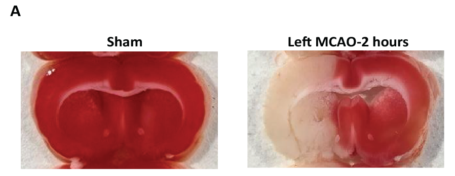
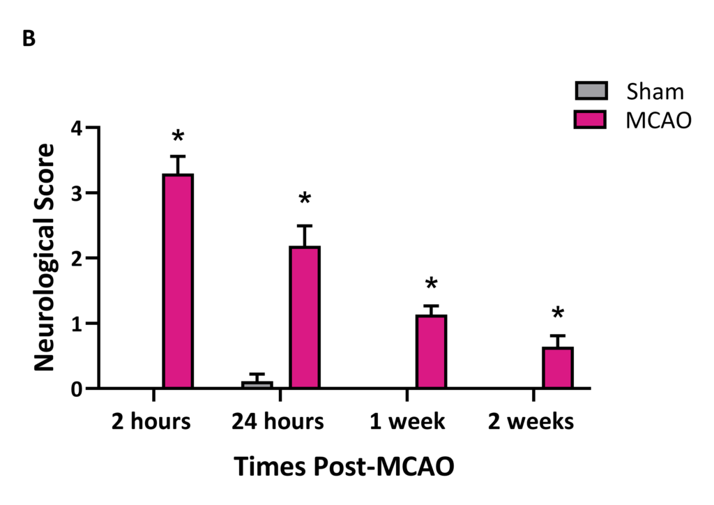
Figure 1 A: TTC staining differentiates viable from infarcted tissue based on metabolic activity. Living tissue reduces TTC to red formazan, while infarcted tissue appears pale. Coronal brain sections collected 24 hours post-reperfusion show infarcts in the striatum and cortex of MCAO rats, whereas brains from sham animals exhibit uniform red staining. B: MCAO rats exhibit increased Neuroscore, reflecting marked neurological impairment. Peak deficits occurred at the end of occlusion, with impairments persisting up to 2 weeks post-stroke.
Sensorimotor Asymmetry
Unilateral brain injury induced by MCAO produces sensorimotor asymmetry that can be measured using the corner and cylinder tests. The corner test evaluates turning bias when rats exit a narrow Plexiglas corner, with consistent directional preference indicating unilateral sensorimotor and postural deficits. The cylinder test assesses forelimb use during vertical exploration, where MCAO rats favor the forelimb ipsilateral to the lesion.
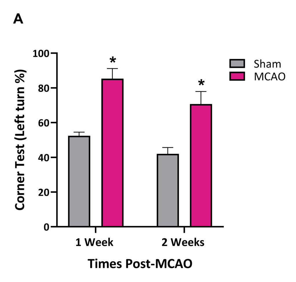
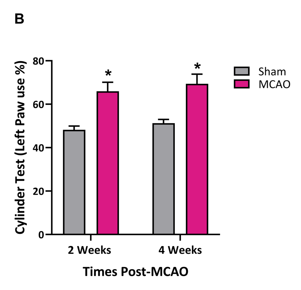
Figure 2: A: MCAO rats show a significant turning bias compared to Sham rats in the Corner test at 1 and 2 weeks post-MCAO, reflecting sensorimotor asymmetry. B: MCAO rats preferentially use the unaffected forelimb in the Cylinder test at 2 and 4 weeks post-MCAO compared to Sham rats.
Balance and Coordination
MCAO rats exhibit balance deficits as measured by their inability to maintain balance on a narrow beam for at least 1 minute. MCAO rats also show impairment in fine motor coordination, specifically, in the placement of the forelimb contralateral to the lesion when crossing a horizontal ladder with irregularly spaced rungs.
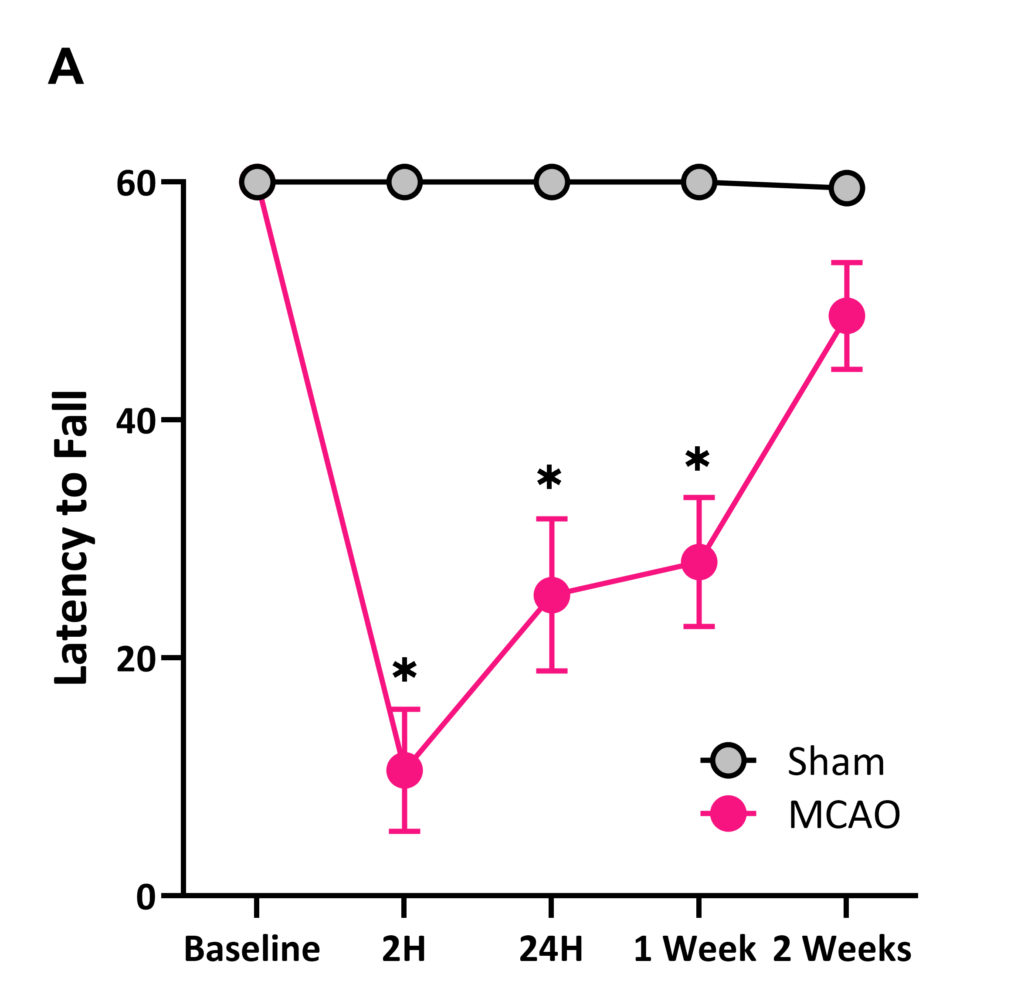
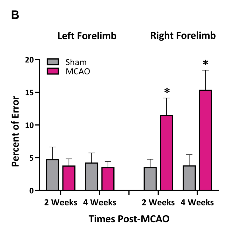
Figure 3 A: MCAO rats display marked balance deficits on the beam balance as measured by the latency to fall when compared to Sham rats. B: MCAO rats show a significant increase in paw misplacement in the contralateral (right) forelimb at 2 and 4 weeks post-stroke.