At PsychoGenics, we offer high-throughput quantitative immunohistochemistry (IHC) and histology services to evaluate and quantify standard and novel biomarkers of disease progression and treatment response. We have expertise in sectioning and staining all tissue types, including Formalin-Fixed Paraffin-Embedded (FFPE) and frozen brain and spinal cord from a variety of species, including rodent disease models and humans, using proprietary and novel antibodies.
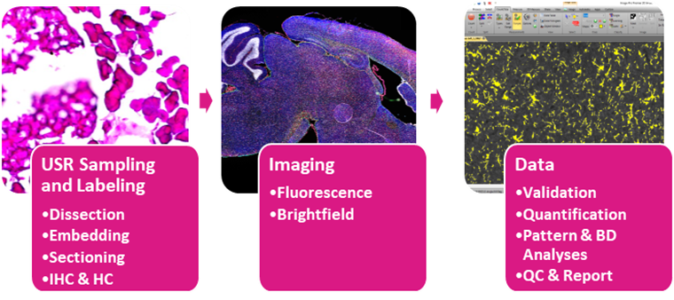
Figure 1.Overview of procedures for tissue processing and data generation, which can be customized for areas of interest and disease models.
Samples and Sectioning
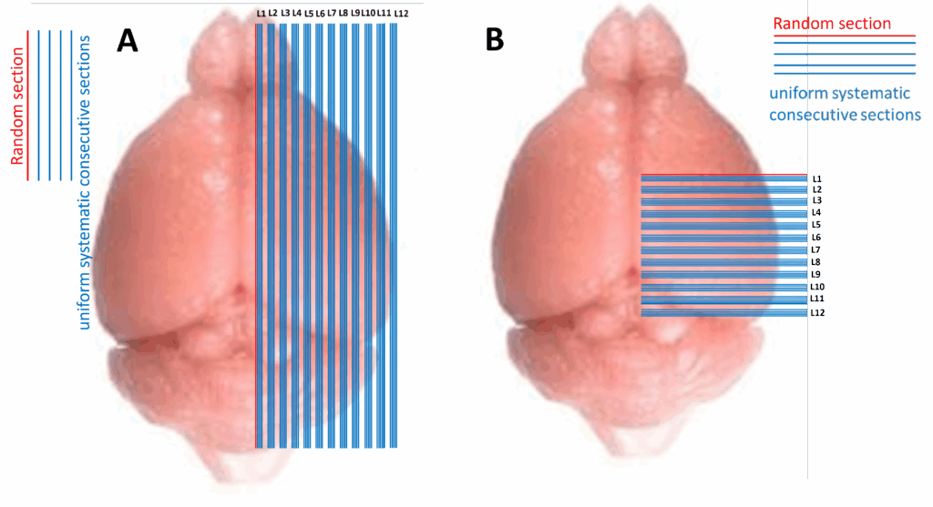
We optimize the preparation of samples for quantitative histology using Uniform Systematic Random (USR) sampling to ensure reproducibility (Figure 2).
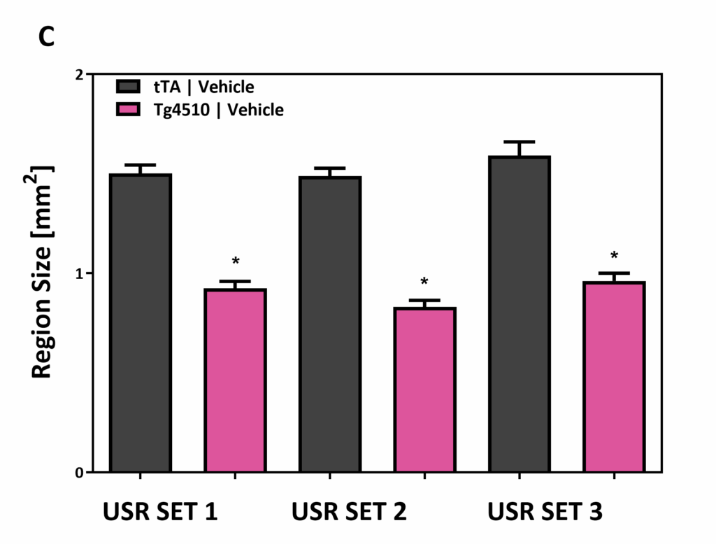
Figure 2. Example of (A) sagittal and (B) coronal USR sectioning. (C) Region size of frontal cortex is significantly reduced in the rTg4510 tauopathy model, as compared to controls. 97% reproducibility of frontal cortex atrophy measurement is obtained from three USR sets from the same animals. Using this approach, sectioning starts with a random section and follows a uniform system to assure reproducibility across studies and over time.
Immunohistochemical Evaluation for Various Disease Models
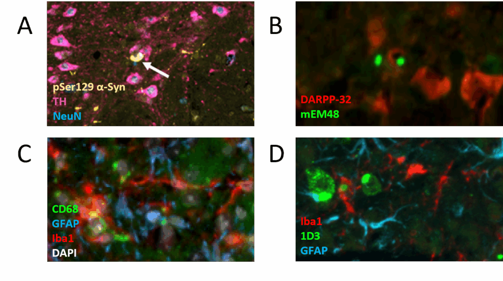
We have extensive experience working with disease models and antibodies (Figure 3) and have validated over 300 primary antibodies to assess many combinations of disease-relevant and morphological markers.
Figure 3. Examples of diverse immunohistochemical markers in rodent disease models. (A) Parkinson’s disease: In fibril-inoculated mice, alpha-synuclein aggregates (pSer129, yellow) form and spread to dopaminergic neurons (TH, pink) in the substantia nigra. (B) Huntington’s disease: R6/1 mice show increased huntingtin aggregation (mEM48, green) and fewer medium spiny neurons (DARPP-32, red) than wild types. (C) Alzheimer’s disease: Pathology in Tg4510 mice is characterized by increased neuroinflammation, including differences in cortical astroglia (GFAP, blue), microglia (Iba1, red), and phagocytic markers (CD68, green). (D) ALS: TDP-43 ΔNLS mice demonstrate higher levels of TDP-43 aggregates (1D3, green) and neuroinflammation (Iba1, red; GFAP, green) in the cortex, as compared to wild types.