The below data was made possible by CMTA, the Charcot-Marie-Tooth Association.
Charcot-Marie-Tooth (CMT) disorders are a family of related disorders that produce progressive distal neuropathies, presenting significant challenges for patients and society. CMT2A is the most common form of CMT Type 2. It is caused by mutation of the MfN2 gene, resulting in axonal degeneration of peripheral nerve, muscle weakness, and atrophy. PsychoGenics, in partnership with the Charcot-Marie-Tooth Association (CMTA), has extensively characterized two rat models of CMT2A expressing human mutations in MfN2, one of which is described below.
Grip Strength Deficits in CMT2A Rats
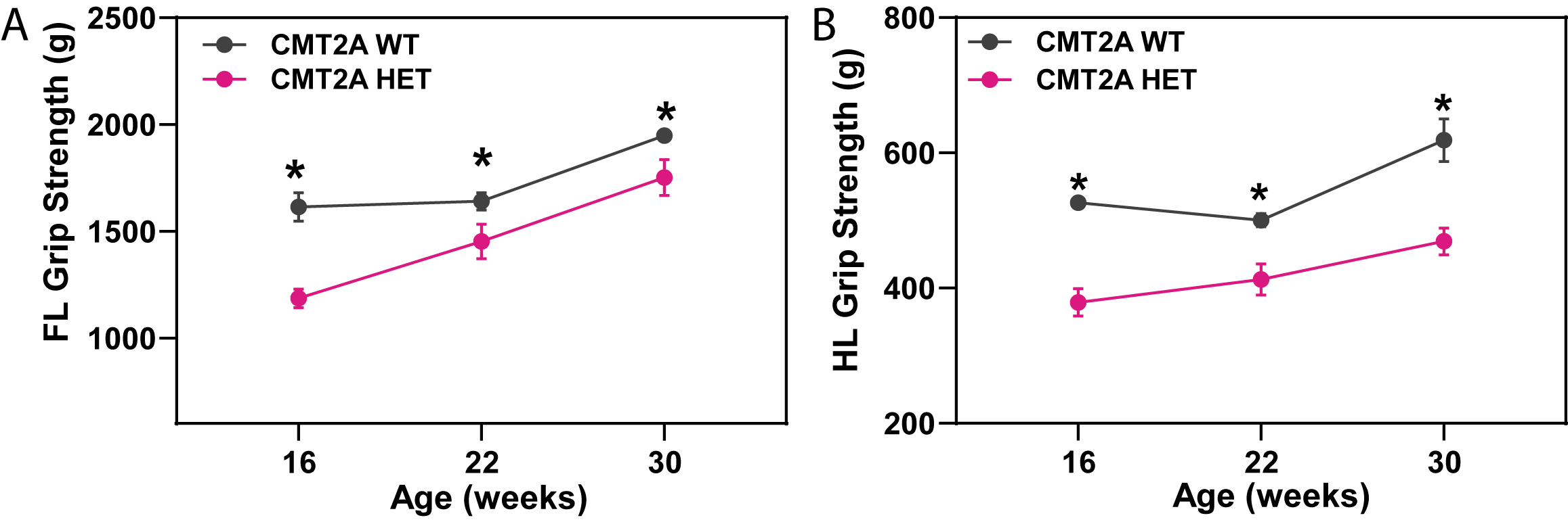
Forelimb (A) and hindlimb (B) grip strength was significantly decreased in male CMT2A HET rats when compared to their WT counterparts.
Gait Analysis
Gait was analyzed using the NeuroCube System, a proprietary automated gait analysis platform. At 30 weeks of age, NeuroCube analysis showed a 90% discrimination in gait features between CMT2A HET and WT rats.
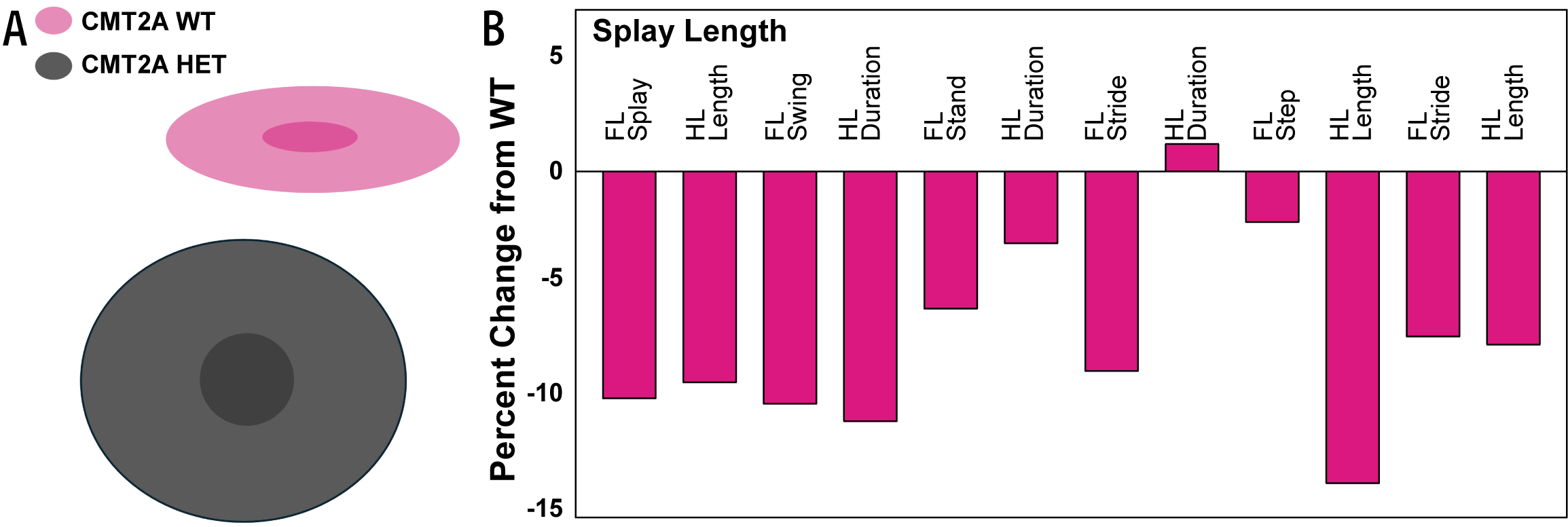
(A) Cloud plots are a way to visualize the difference between the WT and CMT2A HET rats when looking at all gait features (90% discrimination) at 30 weeks of age. (B) Percent changes in gait dynamic and gait geometry features in CMT2A HET rats as compared to WT rats. FL: Forelimb; HL: Hindlimb.
Nerve Conduction Deficits in CMT2A Rats
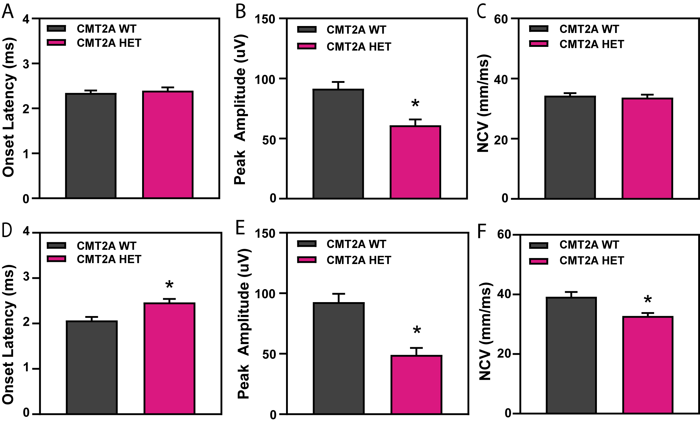
Compound action potential (CAP) at 22 (A-C) and 30 (D-F) weeks of age. CAP responses were obtained from mixed nerve bundles in the tail, comprised of largely sensory axons. Peak amplitude (A&D), which corresponds to the number of functional axons available to be activated, was reduced in the CMT2A HET rats at both ages. Onset latency (B&E), which corresponds to the time from initiation of nerve stimulus to response, was increased in the HET rats at 30 weeks of age, and nerve conduction velocity (C&F) was decreased in the 30-week-old HET rats.
Electron Microscopy in Tibial Nerve
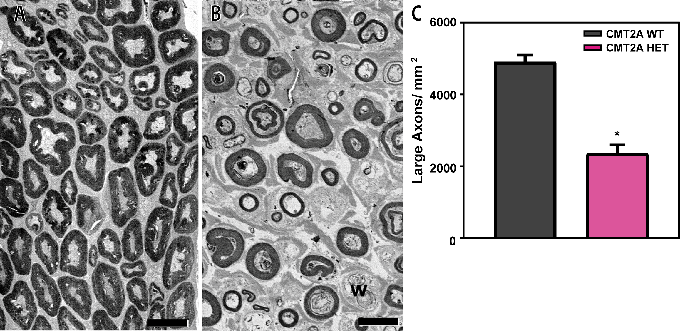
Electron microscopic analysis of tibial nerve from male WT (A) and CMT2A rats (B) at 30 weeks of age. Large motor axons of diameter >5 µm are common, and axoplasm contains intact mitochondria in WT rats (A). In CMT2A HET rats (B), axons were smaller and contained accumulations of disrupted mitochondria and organelles undergoing degeneration. Tibial nerves of CMT2A rats had significantly fewer axons of diameter >5 µm (C). Scale bar 10 µm.
Acknowledgment: Electron microscopic analysis was conducted by Dr. Grahame Kidd at the Cleveland Clinic.
To learn more about preclinical CMT models, contact: Dr. Mark Scheideler, at CMTA, or Dr. Taleen Hanania, at PsychoGenics.