The below data was made possible by CMTA, the Charcot-Marie-Tooth Association.
Charcot-Marie-Tooth (CMT) disorders are a family of related disorders that produce progressive distal neuropathies, presenting significant challenges for patients and society. CMT2E is a form of CMT caused by dominant mutations in the Nefl gene that encodes the neurofilament light protein. Psychogenics, in partnership with the CMT Association has performed extensive characterization of a mouse model of CMT2E.
CMT2E Mice Show Impaired Performance on the Tapered Balance Beam

Male CMT2EA mice take longer to traverse the tapered balance beam at 14 and 20 weeks of age (A) and have a greater number of foot slips (B) compared to WT mice.
Gait Analysis
Gait analysis was conducted using the NeuroCube System, a proprietary automated gait analysis platform. Changes in gait in CMT2E HET rats are observed at 20 weeks of age and become prominent at 26 weeks of age.

(A) Cloud plots are a way to visualize how different the WT and CMT2E HET mice are when looking at all gait features (86% discrimination) at 26 weeks of age. (B) Percent decrease in gait dynamic and gait geometry features in CMT2E HET mice when compared to their WT counterparts. FL: Forelimb; HL: Hindlimb.
Nerve Conduction Deficits in CMT2E Mice
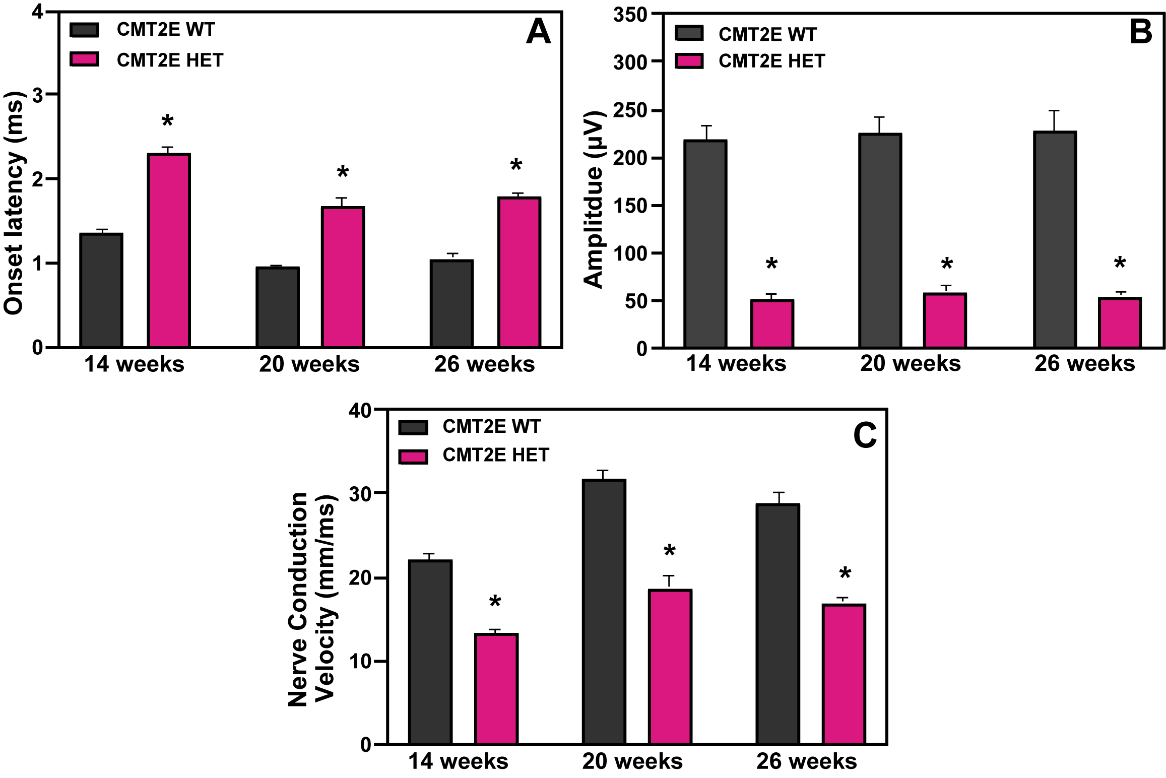
Compound action potential (CAP) at 14, 20 and 26 weeks of age. CAP responses were obtained from mixed nerve bundles in the tail, comprised of largely sensory axons. Onset latency (A), that corresponds to the time from initiation of nerve stimulus to response, was increased in the male CMT2E HET mice at all ages. Peak amplitude (B), corresponding to the number of functional axons available to be activated, was reduced in the male CMT2E HET mice and nerve conduction velocity (C) was also decreased in the HET mice at all ages when compared to their age-matched WT counterparts.
Electron Microscopy in Tibial Nerve
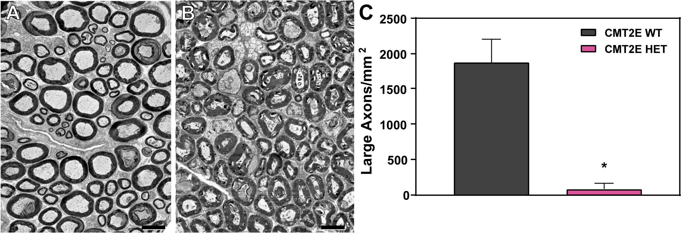
Electron microscopic analysis of tibial nerve from male WT (A) and CMT2E mice (B) at 26 weeks of age. In wild type mice, large motor axons of diameters >3.5 µm are common, and axoplasm contains intact mitochondria and cytoskeleton. In CMT2E mice (B), axons were smaller, and axoplasm contained accumulations of disrupted organelles or flocculent material. Scale bar 5 µm. (C) Tibial nerves from CMTA 2E mice had <5% of axons that were 3 µm or more in diameter, compared with 55% in WT.
Acknowledgment: The CMT2E mouse model was obtained by the CMTA from Dr. R. Lien at Columbia University. Electron microscopic analysis was conducted by Dr. Grahame Kidd at the Cleveland Clinic.
Please also see related work: Lancaster E, Li J, Hanania T, Liem R, Scheideler MA, Scherer SS. Myelinated axons fail to develop properly in a genetically authentic mouse model of Charcot-Marie-Tooth disease type 2E. Exp Neurol. 2018, 308:13-25.